Bowing & Knock Knees

  • Home
  • knocking of knees are commonly seen developmental issues in teenagers, can be treated with minimal invasive surgery if diagnosed on time.

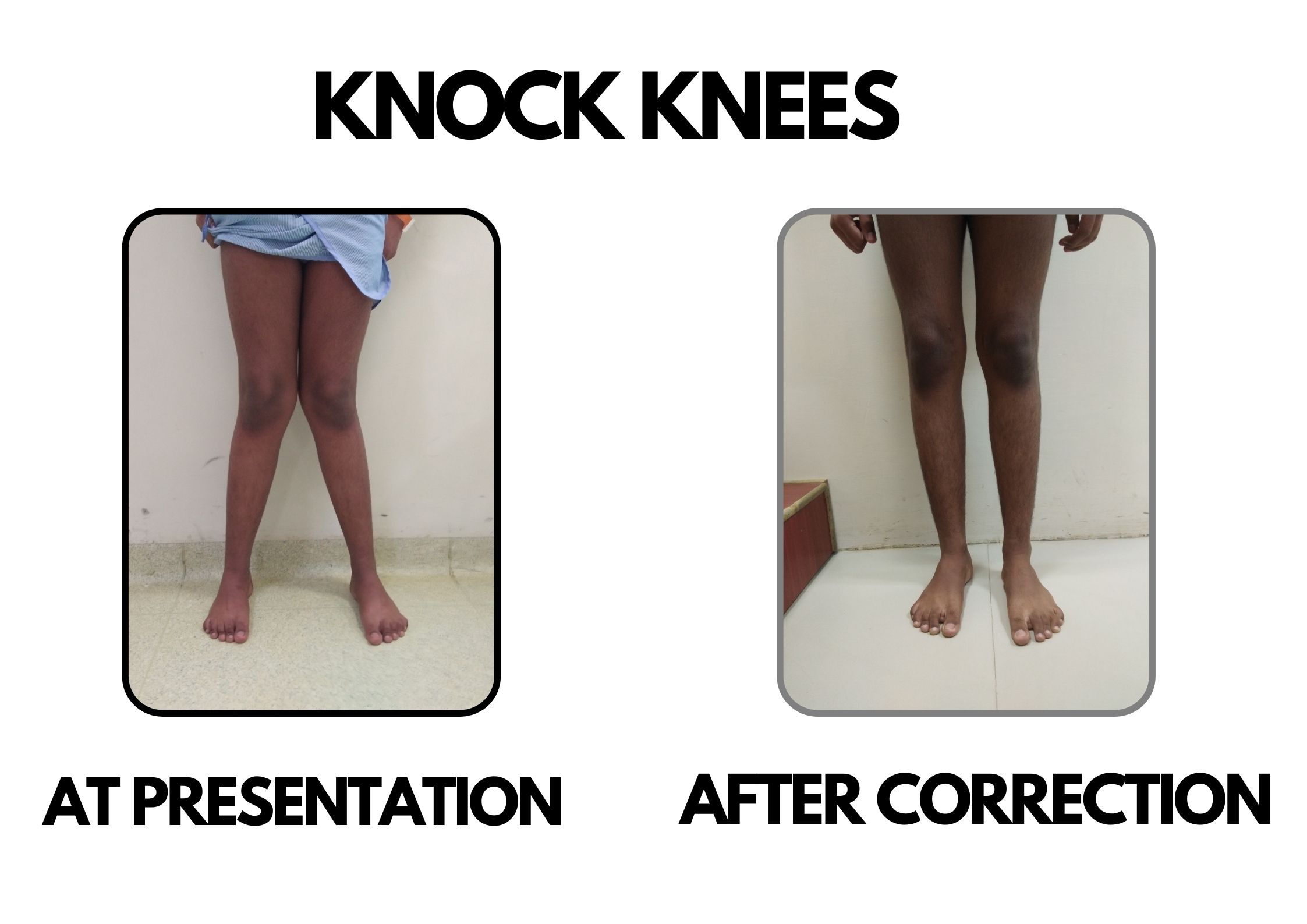
Bowing of legs and knocking of knees are commonly seen developmental issues in toddlers. They can also be seen in teenagers.

In bowing, both the knees go apart and feet come close whereas in knock knees both the knees touch each other and feet go apart. These are variations of the normal biomechanics of the body. All the new born babies have some bowing at the time of birth due to posture of child in the womb. This progressively decreases and children normally grow out of this before 2 years.

However in some conditions this bowing persists or even aggravates and thus needs further investigation in form of standing x rays of both lower limbs to look for alignment and mechanical axis, x rays of both wrists to look for rachitic changes, blood parameters including calcium, phosphorous, vit d and alkaline phosphatase levels.

These investigations can help in differentiating between physiological bowing, blount’s disease (disease of proximal tibial growth plate leading to bowing of knees) and rickets.

After attaning neutral alignment, knocking of knees start developing maximum till 6 years and then decreases till 8 years to attain adult pattern of 5 -7degrees of valgus alignment. Knocking of knees appearing later in childhood warrants investigations including blood investigations for rickets or osteomalacia. Some skeletal dysplasias also can cause knocking of knees.

History and clinical examination involves inquiring about child’s nutrition, growth and development, bowel habits, any family history of skeletal dysplasias, etc. The doctor will have a general physical examination and a detailed examination of the musculoskeletal system including hips, knees and ankles, any limb length discrepancy, ligament laxity, torsional alignment, walking pattern of the child, etc.

Investigations include standing alignment views of both lower limbs, blood investigations to look for changes of rickets.

Treatment depends upon the diagnosis:

  •  In case of rickets, vit d and calcium supplementation by a paediatric orthopaedic surgeon or in association with a paediatric endocrinologist is sufficient. Sequential follow up x rays and blood tests are required to document recovery and improvement in mechanical axis of the lower limbs.

  •  Blount’s disease: It iss due to abnormal physeal growth leading to progressively increasing genu varum. Depending on the classification and grade of proximal physeal involvement, treatment varies from observation to surgical intervention. In cases of adolescent genu varum and valgum, growth modulation is a very good option with predictable results.

Those children who have completed bone growth need corrective osteotomy to rectify the alignment.

Dr. Deepak Khurana

8 Years Experience

MBBS, MS (Orthopaedics) FIPO (Mumbai)

Consultant Paediatric Orthopaedic & Limb reconstruction surgeon.
Testimonials

What Parents Say About Us

Attached Hospitals